

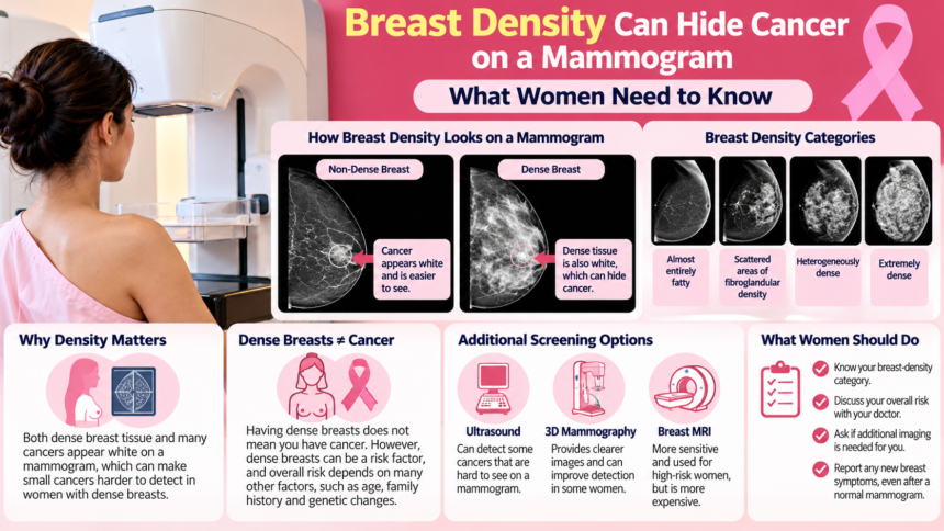
A mammogram can help detect breast cancer before a lump becomes noticeable. However, in women with dense breast tissue, some cancers can be harder to identify because the tissue and many cancerous abnormalities appear white on mammogram images. This overlap can create what doctors call a “masking effect.”
Dense breasts do not mean a woman has breast cancer, and mammograms remain an important screening tool. However, understanding breast density can help women and their doctors make informed decisions about screening and whether additional imaging may be useful.
What Does Having Dense Breasts Mean?
Breast density refers to the proportion of fibrous and glandular tissue compared with fatty tissue in the breasts. Radiologists assess density using mammograms and classify it into four categories:
- Almost entirely fatty: The breasts contain mostly fatty tissue.
- Scattered areas of fibroglandular density: Some areas contain dense tissue, but most of the breast is fatty.
- Heterogeneously dense: A substantial amount of dense tissue is present.
- Extremely dense: Most of the breast consists of dense tissue.
Breast density cannot be reliably determined by looking at or feeling the breasts. It is assessed through mammography.
Density can change throughout a woman’s life. It is generally higher in younger women and often decreases after menopause. Hormonal factors, pregnancy, breastfeeding and certain medications can also influence breast composition.
Why Can Dense Breast Tissue Make Cancer Harder to Detect?
On a mammogram, fatty tissue usually appears dark, while fibrous and glandular tissue appears white. Many breast cancers also appear as white areas, which can make them difficult to distinguish from the surrounding tissue in dense breasts.
In breasts containing more fatty tissue, an abnormality may stand out more clearly against the darker background. In heterogeneously or extremely dense breasts, overlapping white tissue can obscure a small cancer and reduce the sensitivity of mammography.
Dense breast tissue can also make screening results more difficult to interpret. Some women may be called back for additional imaging or undergo a biopsy to investigate findings that ultimately turn out to be benign.
Do Dense Breasts Increase Breast Cancer Risk?
Breast density and breast cancer are related, but they are not the same thing. Having dense breasts does not mean a woman has cancer. However, breast density is an independent risk factor, and the risk is generally higher in women with extremely dense breasts.
A woman’s overall risk also depends on other factors, including:
- Age and family history of breast cancer.
- Previous breast cancer or certain atypical findings on breast biopsy.
- Inherited genetic variants, including BRCA1 and BRCA2.
- Previous significant exposure to radiation.
For this reason, two women with the same breast-density category may need different screening approaches. A woman with dense breasts and a strong family history may have a different overall risk from someone with dense breasts but no other major risk factors.
Breast cancer is a significant health concern in India. Estimates from GLOBOCAN 2022 and India’s National Cancer Registry Programme differ because they use different estimation and registration methods, but both indicate a substantial burden of disease among women.
What Additional Screening Options Are Available?
Depending on breast density and overall cancer risk, doctors may consider additional imaging alongside mammography.
3D mammography: Also called digital breast tomosynthesis, this technique takes multiple low-dose X-ray images from different angles to create thin image sections of the breast. It can reduce the effects of overlapping tissue and improve cancer detection in some women.
Breast ultrasound: Ultrasound uses sound waves to examine breast tissue and may identify some abnormalities that are difficult to see on a mammogram. However, it can also detect benign findings that lead to further tests or unnecessary biopsies.
Breast MRI: Magnetic resonance imaging is particularly useful for certain women at high risk, including some women with inherited genetic variants or a calculated lifetime breast cancer risk of 20% or more. MRI is more sensitive than mammography but can be expensive, requires contrast in standard screening protocols and may reveal findings that need further investigation.
Additional imaging is not automatically necessary for every woman with dense breasts. Decisions should take account of age, density, family history, overall risk, potential benefits, false-positive results, cost and availability.
When Should Women Start Screening?
Screening recommendations can differ between organisations and may change as evidence develops. Indian guidelines include age-based and risk-based approaches, with recommendations for mammography beginning at age 40 in certain guidance.
The appropriate schedule depends on the applicable guideline, personal risk factors and advice from a healthcare professional. Women with a strong family history or other significant risk factors may need an individualised screening plan.
If a mammogram report identifies dense breasts, consider asking your doctor:
- What is my breast-density category?
- Does the density make cancer harder to detect on my mammogram?
- What is my overall breast cancer risk?
- Would 3D mammography, ultrasound or MRI provide a meaningful benefit?
- What are the possible disadvantages of additional screening?
Why Breast Symptoms Should Never Be Ignored
A normal mammogram does not rule out every breast cancer, particularly when breast tissue is dense. A new lump, unusual nipple discharge, changes to the skin or nipple, or another persistent breast change should be assessed by a healthcare professional, even if a recent mammogram was normal.